Brain Tumor
A brain tumor is an abnormal growth of cells within the brain, which can be cancerous(malignant) or non-cancerous (benign). It is defined as any intracranial tumor created by abnormal and uncontrolled cell division, normally either in the brain itself (neurons, glial cells (astrocytes,oligodendrocytes, ependymal cells, myelin-producing Schwann cells), lymphatic tissue, blood vessels), in the cranial nerves, in the brain envelopes (meninges), skull, pituitary and pineal gland, or spread from cancers primarily located in other organs (metastatic tumors).
Primary (true) brain tumors are commonly located in the posterior cranial fossa in children and in the anterior two-thirds of the cerebral hemispheres in adult, although they can affect any part of the brain.
In the United States in the year 2005, it was estimated there were 43,800 new cases of brain tumors (Central Brain Tumor Registry of the United States, Primary Brain Tumors in the United States, Statistical Report, 2005–2006),[1] which accounted for 1.4 percent of all cancers, 2.4 percent of all cancer deaths,[2] and 20–25 percent of pediatric cancers.[2][3] Ultimately, it is estimated there are 13,000 deaths per year in the United States alone as a result of brain tumors.[1]
Contents[hide] |
[edit]Causes
Metastatic cancers are far more common than primary tumors of the brain and spinal cord.
Apart from exposure to vinyl chloride or ionizing radiation, there are no known environmental factors associated with brain tumors. Mutations and deletions of so-called tumor suppressor genes are thought to be the cause of some forms of brain tumors. Patients with various inherited diseases, such as Von Hippel-Lindau syndrome, multiple endocrine neoplasia, neurofibromatosis type 2 are at high risk of developing brain tumors. It is alleged that mobile phones/cell phones might be a cause of brain tumors, according to one report.[4] (see Mobile phone radiation and health) There is an association of brain tumor incidence and malaria, suggesting that the anopheles mosquito, the carrier of malaria, might transmit a virus or other agent that could cause a brain tumor.[5] Malignant brain tumor incidence and Alzheimer's disease prevalence are associated in 19 US states. The two diseases may share a common cause, possibly inflammation.[6]
[edit]Signs and symptoms
Symptoms of brain tumors may depend on two factors: tumor size (volume) and tumor location. The time point of symptom onset in the course of disease correlates in many cases with the nature of the tumor ("benign", i.e. slow-growing/late symptom onset, or malignant, fast growing/early symptom onset) is a frequent reason for seeking medical attention in brain tumor cases..
Large tumors or tumors with extensive perifocal swelling edema inevitably lead to elevated intracranial pressure (intracranial hypertension), which translates clinically into headaches, vomiting (sometimes without nausea), altered state of consciousness (somnolence, coma), dilatation of the pupil on the side of the lesion (anisocoria), papilledema (prominent optic disc at the funduscopic eye examination). However, even small tumors obstructing the passage of cerebrospinal fluid (CSF) may cause early signs of increased intracranial pressure. Increasedintracranial pressure may result in herniation (i.e. displacement) of certain parts of the brain, such as the cerebellar tonsils or the temporaluncus, resulting in lethal brainstem compression. In young children, elevated intracranial pressure may cause an increase in the diameter of theskull and bulging of the fontanelles.
Depending on the tumor location and the damage it may have caused to surrounding brain structures, either through compression or infiltration, any type of focal neurologic symptoms may occur, such as cognitive and behavioral impairment, personality changes, hemiparesis,hypoesthesia, aphasia, ataxia, visual field impairment, facial paralysis, double vision, tremor etc. These symptoms are not specific for brain tumors—they may be caused by a large variety of neurologic conditions (e.g. stroke, traumatic brain injury). What counts, however, is the location of the lesion and the functional systems (e.g. motor, sensory, visual, etc.) it affects.
A bilateral temporal visual field defect (bitemporal hemianopia—due to compression of the optic chiasm), often associated with endocrine disfunction—either hypopituitarism or hyperproduction of pituitary hormones and hyperprolactinemia is suggestive of a pituitary tumor.
[edit]Types of brain tumors
- Glioblastoma multiforme
- Medulloblastoma
- Astrocytoma
- CNS lymphoma
- Brainstem glioma
- Germinoma
- Meningioma
- Oligodendroglioma
- Schwannoma
- Craniopharyngioma
- Ependymoma
- Mixed gliomas
- Brain metastasis[7]
[edit]Diagnosis
Although there is no specific clinical symptom or sign for brain tumors, slowly progressive focal neurologic signs and signs of elevated intracranial pressure, as well as epilepsy in a patient with a negative history for epilepsy should raise red flags. However, a sudden onset of symptoms, such as an epileptic seizure in a patient with no prior history of epilepsy, suddenintracranial hypertension (this may be due to bleeding within the tumor, brain swelling or obstruction of cerebrospinal fluid's passage) is also possible.
Glioblastoma multiforme and anaplastic astrocytoma have been associated in case reports onPubMed[who?] with the genetic acute hepatic porphyrias (PCT, AIP, HCP and VP), including positive testing associated with drug refractory seizures. Unexplained complications associated with drug treatments with these tumors should alert physicians to an undiagnosed neurological porphyria.
Imaging plays a central role in the diagnosis of brain tumors. Early imaging methods—invasive and sometimes dangerous—such as pneumoencephalography and cerebral angiography, have been abandoned in recent times in favor of non-invasive, high-resolution modalities, such as computed tomography (CT) and especially magnetic resonance imaging (MRI). Benign brain tumors often show up as hypodense (darker than brain tissue) mass lesions on cranial CT-scans. On MRI, they appear either hypo- (darker than brain tissue) or isointense (same intensity as brain tissue) on T1-weighted scans, or hyperintense (brighter than brain tissue) on T2-weighted MRI, although the appearance is variable. Perifocal edema also appears hyperintense on T2-weighted MRI. Contrast agent uptake, sometimes in characteristic patterns, can be demonstrated on either CT or MRI-scans in most malignant primary and metastatic brain tumors. This is because these tumors disrupt the normal functioning of the blood-brain barrier and lead to an increase in its permeability. However it is not possible to diagnose high versus low grame gliomas based on enhancement pattern alone.
Electrophysiological exams, such as electroencephalography (EEG) play a marginal role in the diagnosis of brain tumors.
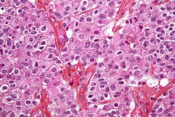
The definitive diagnosis of brain tumor can only be confirmed by histological examination of tumor tissue samples obtained either by means of brain biopsy or open surgery. The histological examination is essential for determining the appropriate treatment and the correct prognosis. This examination, performed by a pathologist, typically has three stages: interoperative examination of fresh tissue, preliminary microscopic examination of prepared tissues, and followup examination of prepared tissues after immunohistochemical staining or genetic analysis.
Another possible diagnosis would be neurofibromatosis which can be in type one or type two.


0 Comments:
Post a Comment
<< Home